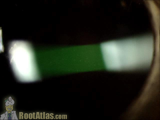
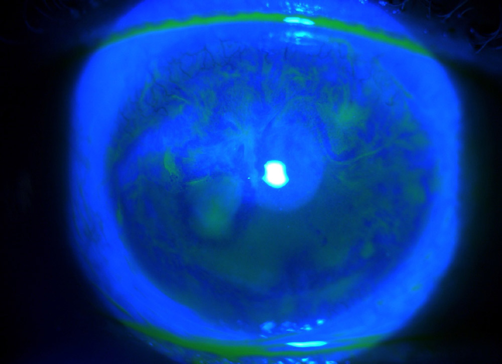
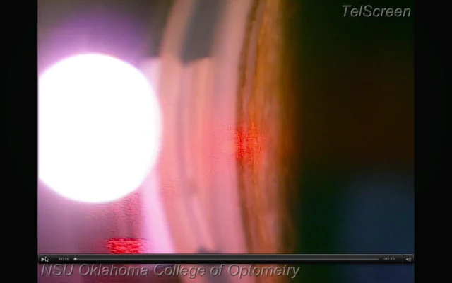
This video demonstrates what cell and flare look like under the slit-lamp microscope. “Cell” is the individual inflammatory cells while “flare” is the foggy appearance given by protein that has leaked from inflamed blood vessels. This finding is commonly seen with uveitis, iritis, and after surgery … and actually seeing it can be challenging for
3 in 1 Phone Camera Lens - 0.65x Super wide Angle Lens + 15x MACRO Lens + 230 degree fisheye lens, Meet all you demand on photography. Easy for you to

Criacr Phone Camera Lens, 230° Fisheye Lens, 15X Macro Lens, 0.65X Wide Angle Lens, Clip-On 3 in 1 Cell Phone Lens for Live Video, Compatible with
Monday Back to Basics

Introduction to Oculoplastics (with Dr. Andrea Tooley) by The Lens Pod

Tugas Modul Gangguan Mata Tutor 10
How vaccines may affect the cornea - EyeWorld

Cells & flare in the anterior chamber

Woche 10 - mariaundleaunterwegss Webseite!

Aqueous flare and cells in Fuchs syndrome
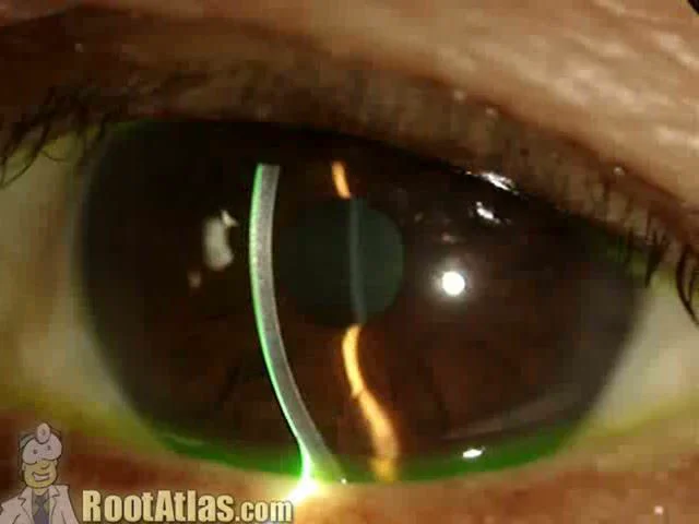
Cell and flare in the eye (Video)
Get Laser Focused on the Appropriate Glaucoma Treatment
Iris Beam's Instagram, Twitter & Facebook on IDCrawl

Sun
Slit lamp examination showing anterior chamber cells and flare

South Alabama Emergency Medicine (@SouthEmergency) / X